Naples Heart Rhythm Specialists, P.A.
Kenneth W. Plunkitt, M.D., Founder
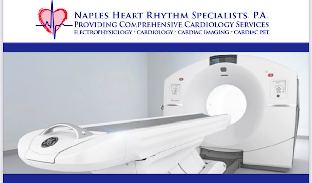
 Naples Heart Rhythm Specialists, P.A. is proud to announce that we now have one of the most advanced cardiac PET machines in the country. In addition to having state of the art PET equipment, our cardiologists are specifically trained in myocardial perfusion nuclear imaging.
What is a cardiac PET scan?
Cardiac positron emission tomography (PET) is a noninvasive diagnostic heart test that uses radionuclides, which are radioactive tracers, to produce unrivaled 3D perfusion and function scans of the heart by evaluating myocardial metabolism.
The radionuclide tracer Rubidium (Rb-82), is rapidly absorbed into the myocardium as it is a potassium analog and actively participates in sodium/potassium exchange pumps. The scanner contains a Gamma ray detection ring that absorbs Gamma rays from a proton annihilation reaction. This event creates a coincidence event in which gamma rays strike the ring in a single vector on opposite sides of the ring. The computer converts the electronic signals into images.
Why would a patient need a cardiac PET scan?
The team at NHRS may recommend a cardiac PET scan to diagnose coronary artery disease, myocardial ischemia, previous myocardial infarction or myocardial viability.
A cardiac PET scan shows myocardial metabolism: therefore, it can also be used to calculate noninvasive coronary flow. Comparisons of rest and stress flows can be a very useful diagnostic tool that can determine small vessel perfusion disease that cannot be evaluated with any other noninvasive imaging modality.
What can be expected during a cardiac PET scan?
Patient will have an IV started and be attached to 12 lead EKG. An initial CT scout scan is followed by a six-minute rest myocardial scan. Once rest scans are processed, a stress agent such as regadenoson (Lexiscan) is infused over a one-minute protocol followed by a six-minute myocardial stress scan. The EKG is assessed for possible ischemic changes. The test is less than 40 minutes in duration.
Is a cardiac PET scan safe?
Compared to a traditional SPECT stress testing, the half-life of Rubidium is 70-80 seconds as opposed to 6 – 70 + hour half-life of traditional SPECT nuclear tracers. The radiation that is used is minute compared to standard CT scans, traditional nuclear SPECT scans, and cardiac catheterization.
In summary, Cardiac PET is faster, safer, non-invasive, more accurate than traditional myocardial perfusion exams, and provides assessment of myocardial blood flow that is normally only available with cardiac catheterization with a flow wire placed in the coronaries themselves.